3-D printed cancer cells to mimic tumours for drug research
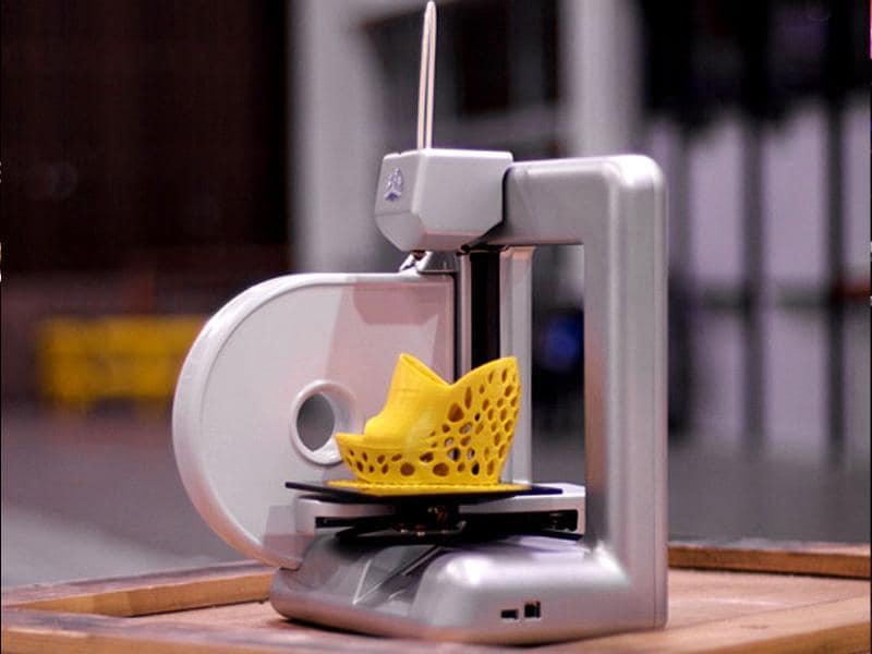
Researchers have successfully created a 3D model of a cancerous tumour using a 3D printer, an advance that can be used to test the efficacy and safety of new cancer drugs and therapies.
| Updated on: Apr 12 2014, 11:19 IST
Researchers have successfully created a 3D model of a cancerous tumour using a 3D printer, an advance that can be used to test the efficacy and safety of new cancer drugs and therapies.
The model, which consists of a scaffold of fibrous proteins coated in cervical cancer cells, has provided a realistic 3D representation of a tumour's environment and could help in the discovery of new drugs and cast new light on how tumours develop, grow and spread throughout the body.
The model consists of a grid structure, 10 mm in width and length, made from gelatin, alginate and fibrin, which recreates the fibrous proteins that make up the extracellular matrix of a tumour.
The grid structure is coated in Hela cells - a unique, 'immortal' cell line that was originally derived from a cervical cancer patient in 1951.
Although the most effective way of studying tumours is to do so in a clinical trial, ethical and safety limitations make it difficult for these types of studies to be carried out on a wide scale.
With the advent of 3D printing, it is now possible to provide a more realistic representation of the environment surrounding a tumour, which the researchers have demonstrated in this study by comparing results from their 3D model with results from a 2D model.
In addition to testing if the cells remained viable, or alive, after printing, the researchers also examined how the cells proliferated, how they expressed a specific set of proteins, and how resistant they were to anti-cancer drugs.
The proteins studied were part of the MMP protein family. These proteins are used by cancer cells to break through their surrounding matrix and help tumours to spread.
Resistance to anti-cancer drugs, which was also studied, is a good indicator of tumour malignancy.
The results revealed that 90 per cent of the cancer cells remained viable after the printing process.
'We have provided a scalable and versatile 3D cancer model that shows a greater resemblance to natural cancer than 2D cultured cancer cells,' said Professor Wei Sun, from Tsinghua University, China, and Drexel University, US.
The study was published the journal Biofabrication.
Catch all the Latest Tech News, Mobile News, Laptop News, Gaming news, Wearables News , How To News, also keep up with us on Whatsapp channel,Twitter, Facebook, Google News, and Instagram. For our latest videos, subscribe to our YouTube channel.